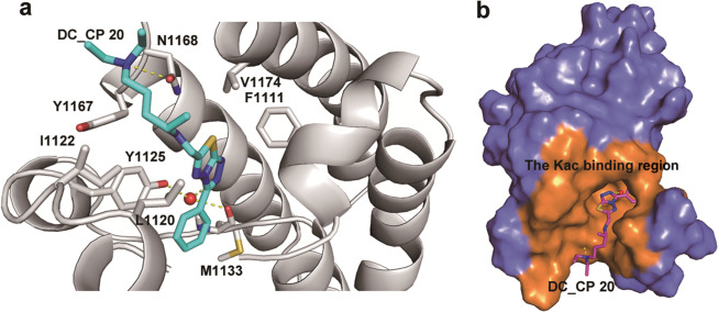
Fig. 3.
Putative binding mode between DC_CP20 and CBP BrD. a A close up view of the interaction between CBP BrD and DC_CP20. The ligand and interacting residues are shown as sticks; water molecules are shown as spheres; hydrogen bonds are indicated by yellow dotted lines. b The Kac-binding region of CBP BrD occupied by DC_CP20

