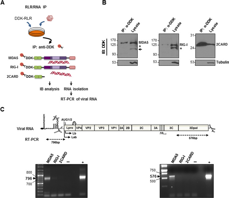
Fig. 1. Isolation of RLR/RNA complexes in FMDV-infected cells.
a Schematic representation of the procedure for viral RNA pulldown. b IBRS-2 cells were transfected with plasmids encoding DDK-tagged -MDA5, -RIG-I or -2CARD (1 µg/106 cells) and infected 24 h later with FMDV O1K isolate at an MOI of 5. Cells were lysed 5 h after infection and lysates were subjected to IP and analyzed by immunoblot with an anti-DDK monoclonal antibody. A faster migration form of MDA5 (about 95 kDa) bearing the N-terminal tag is indicated with an arrow. Asterisk denotes MDA5 and RIG-I forms with slightly faster migration commonly observed in cells expressing the RLRs. c RNA was extracted from the IP fractions and analyzed by RT-PCR with two sets of primers for amplification of the indicated 796-bp 5´- and 576-bp 3´- terminal regions of the viral RNA. The specific IP fraction (MDA5, RIG-I or 2CARD) corresponding to each lane is indicated. Negative and positive controls were included in the RT-PCR assays (water and in vitro transcribed FMDV O1K RNA, respectively).

