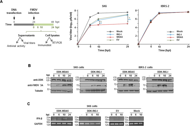
Fig. 3. Effect of RIG-I and MDA5 expression on FMDV growth.
SK6 or IBRS-2 cells (1 × 10⁶) were transfected with 2 µg of plasmids encoding DDK-MDA5, DDK-RIG-I, an empty vector (EV) or mock-transfected. Cells were infected 24 h later with FMDV O1K isolate at an MOI of 1. Supernatants were collected and cells lysed at the indicated times after infection. a A schematic representation of the experiment is shown. Viral titers in supernatants collected from SK6 or IBRS-2 cells were determined by plaque assay. Data are mean ± SD of duplicates of two independent experiments. Significant differences using the Student´s t test between viral titers in EV- or RIG-I-transfected and MDA5-transfected SK6 cells (green and blue asterisks, respectively) are indicated (*p < 0.05; **p < 0.01). b Cell lysates were analyzed by immunoblot for the DDK-tagged proteins and viral 3A non-structural protein. Tubulin was used for normalization. c RT-PCR amplification of IFN-β and GAPDH mRNAs from RNA extracted from SK6 cells transfected as specified or mock-transfected and lysed at the indicated times after infection. Due to extensive CPE at 24 hpi, RNA extraction from the few remaining attached cells was only possible for MDA5-transfected monolayers at that time point. GAPDH levels were used as loading control.

