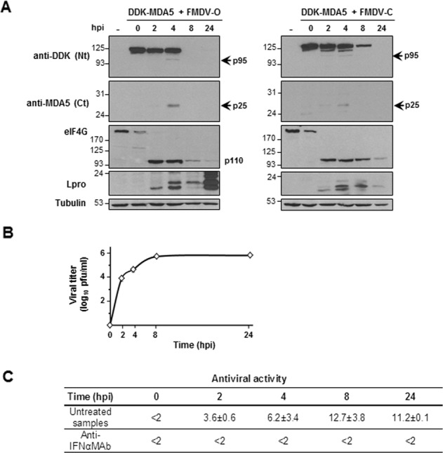
Fig. 4. FMDV infection induces MDA5 cleavage.

Swine SK6 cells were mock-transfected or transfected with 2 µg of a plasmid encoding DDK-MDA5 and 24 h later infected with type-O or type-C FMDV at an MOI of 5. Cells were lysed at different times after infection. a Lysates were analyzed by immunoblot for detection of the indicated proteins using the specified antibodies. Arrows indicate the N- and C-terminal cleavage products of MDA5. The 110-kDa cleavage product of eIF4G is also depicted. b Viral titers in supernatants collected from cells analyzed in a (infected with type-O virus) were determined by plaque assay on IBRS-2 cells. Data as mean of triplicates ± SD. c Antiviral activity in supernatants from cells analyzed in a (infected with type-O virus) is expressed as the reciprocal of the highest dilution of the supernatant needed to reduce the number of VSV plaques on IBRS-2 cells by 50%. When indicated, supernatants were previously treated with a monoclonal antibody anti-swine IFN-α. Data are average of three independent assays ± SD.
