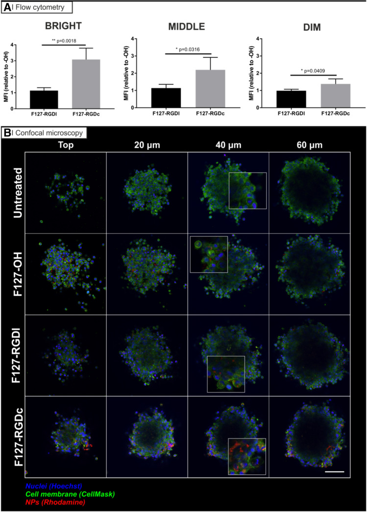
Figure 6.
Penetration of rhodamine-labelled PLGA nanoparticles (red) prepared from Pluronic F127-OH, Pluronic F127-RGDl or Pluronic F127-RGDc (0 and 100% RGD/OH mixtures) in U87MG multicellular spheroids. (A) Flow cytometry analysis: spheroids of about 400 µm in diameter were treated for 24 h with the three different nanoparticle formulations in cell culture media (0.5 mg/mL) at 37 ˚C, and then incubated with Hoechst (to differentiate between the edge and the inner part of the spheroid) for 10 min before disaggregation with trypsin/EDTA and flow cytometry processing. The graphs are representative of 3 independent experiments. Statistical analysis (Unpaired t-test), n = 3. (B) Confocal microscopy analysis: spheroids of about 250 µm in diameter were treated for 24 h with the three different nanoparticle formulations in cell culture media (0.5 mg/mL) at 37 ˚C. They were then incubated with Hoechst for 10 min before confocal microscopy analysis. Images are representative of 3 independent experiments. Nuclei: Hoechst stain (blue); Cell membranes: CellMask (green). Scale bar = 50 μm.

