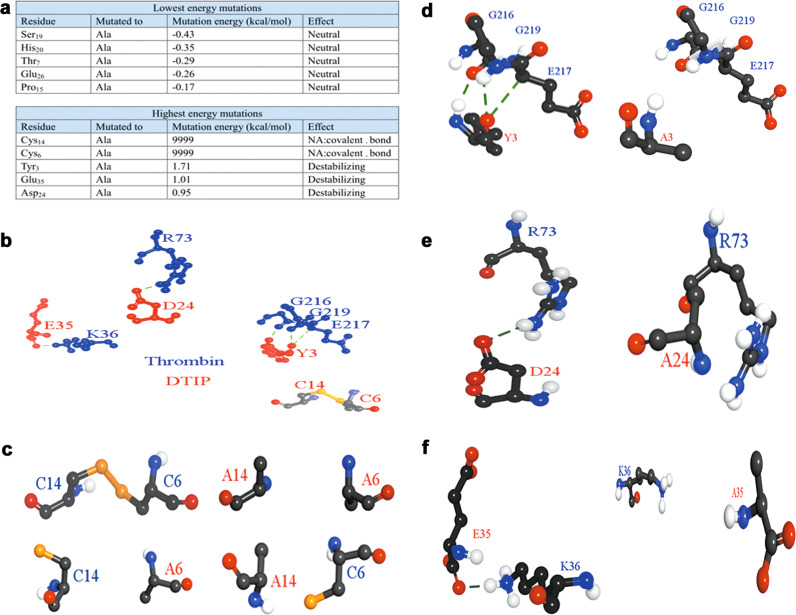
Fig. 5. Alanine scanning and the function bonds of five amino acid residues of DTIP.
a The result of alanine scanning. Amino acid residues with lower energy mutations (up); amino acid residues with higher energy mutations (down). b Hydrogen bonds between five amino acid residues of DTIP (red) and thrombin (blue); disulfide bond between Cys6 and Cys14 of DTIP. c–f Comparison of structures before and after mutation.

