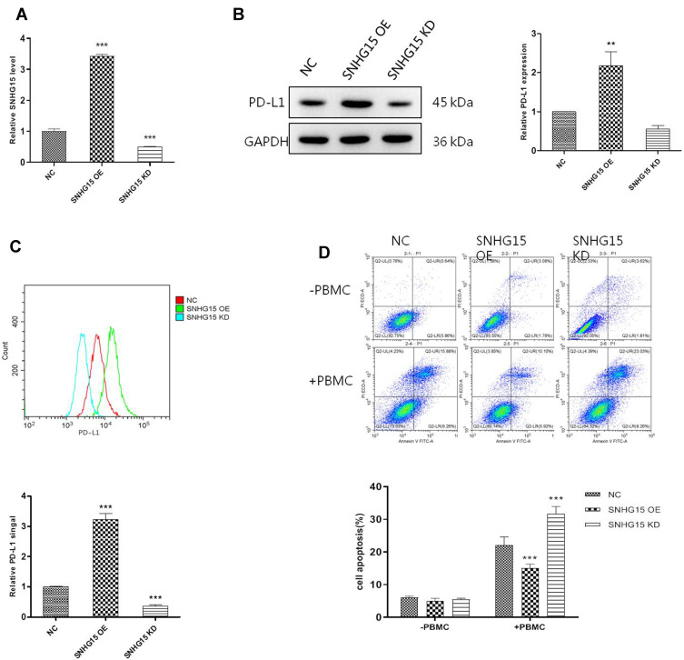
Figure 2.
SNHG15 promoted the expression of PD-L1 and inhibited cell apoptosis. (A) The expression of SNHG15 in HGC-27 cells after different treatments. (B) Whole cell PD-L1 expression was detected with Western Blot. (C) Surface PD-L1 expression was detected with flow cytometry. (D) Cell apoptosis was detected with flow cytometry after HGC-27 cells was transfected with SNHG15 overexpression plasmid or siRNA and incubated with PBMC for another 24 hours. Mean values (±SD) were calculated from three replications. **P < 0.01, ***P < 0.001.

