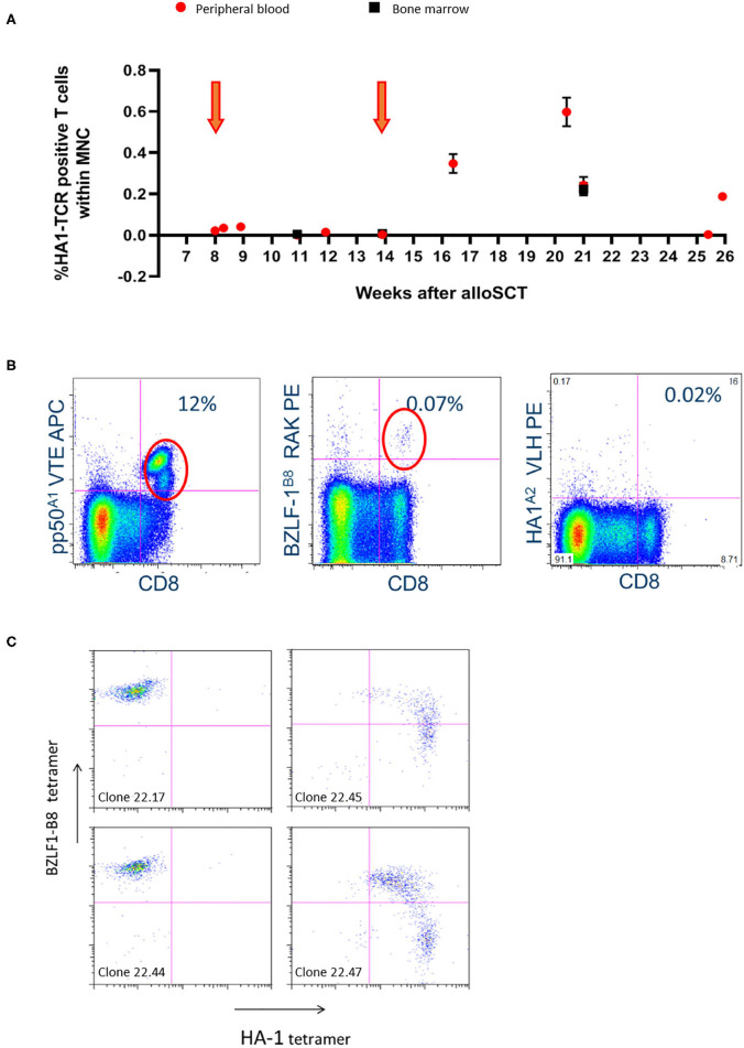
Figure 1.
Significant in-vivo persistence of HA-1H TCR-transduced T cells could be observed during follow-up with evidence of expansion after the second infusion in patient 001. (A) Vector-specific qPCR analysis was performed on peripheral blood and bone marrow samples at indicated time-points. Six weeks after the second infusion the highest peak of HA-1H TCR-transduced CMV or EBV-specific T cells peripheral blood and bone marrow samples was detected. Orange arrows illustrate infusion of HA-1h TCR modified T cells. (B) Facs analysis was performed on peripheral blood sample 6 weeks after infusion of the second cell line. Low numbers of EBV-specific T cells were observed (0.07%), and high frequencies of CMV-specific T cells were found including the infused CMV-pp50-A1-VTE specificity (12%). (C) T cells were isolated from PBMCs 6 weeks after second infusion using EBV-BZLF1-B8-RAK tetramers, single cell sorted and expanded, and tested after 14 days with the different pMHC-tetramers indicated. 50% of the EBV-BZLF1-B8-RAK-specific T-cell clones expressed the HA-1H TCR as measured by pMHC-tetramer analysis.

