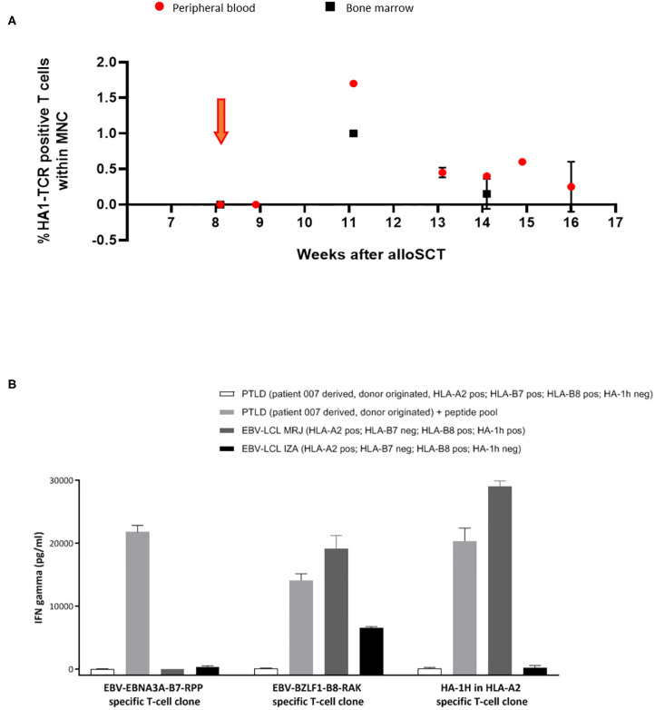
Figure 3.
Significant expansion and persistence of HA-1H TCR-transduced T cells could be documented in peripheral blood and bone marrow samples of patient 007. (A) Vector -specific qPCR analysis was performed on peripheral blood and bone marrow samples at indicated time-points. Orange arrow illustrates infusion of HA-1H TCR-modified T cells. (B) EBNA-3A-B7 and BZLF1-B8 and HA1H specific T cells were stimulated with patient 007 PTLD (SLC PTLD) that was not loaded or loaded with pool of the 2 EBV peptides, LCL-MRJ (HLA-A2+, B7-, B8+, HA-1H+, BZLF1+), and LCL-IZA (HLA-A2+, B7-, B8+, HA-1H-, BZLF1+). PTLD of patient 007 was not recognized by EBV-specific T-cell populations unless they were exogenously loaded with EBV-specific peptides. This PTLD was also not recognized by HA-1H-specific T cells, because the PTLD was donor-derived and therefore HA-1H negative. PTLD sample consisted of monoclonal B cells after CD19 enrichment of PBMNC.

