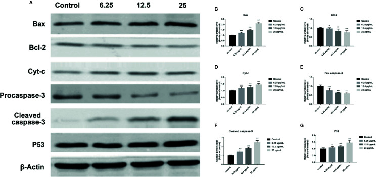
Figure 4.
GA affected the expression of mitochondrial dysfunction related apoptosis proteins in T24 cells. (A–G) Representative western blot images and quantitative analyses of apoptosis-related proteins, including Bax (A, B), Bcl-2 (A, C), Cyt-c (A, D), Pro-caspase-3 (A, E), Cleaved caspase-3 (A, F), and P53 (A, G), in T24 cells with different concentrations of GA treatment. * p < 0.05, ** p < 0.01, *** p < 0.001, compared to the control group. β-Actin was served as the internal reference.

