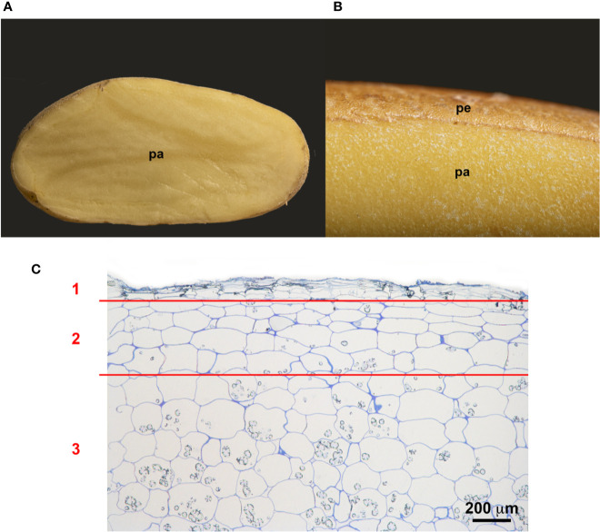
Figure 2.
Structure of the potato periderm. (A) Longitudinal section of a potato tuber. (B) Close view of the potato periderm (pe) and parenchyma (pa). (C) Microscopic semithin section of the suberized phellem (1), the starch-depleted phelloderm (2) and the starch-containing storage parenchymal cells (3). Bar corresponds to 200 µm.

