Fig. 2.
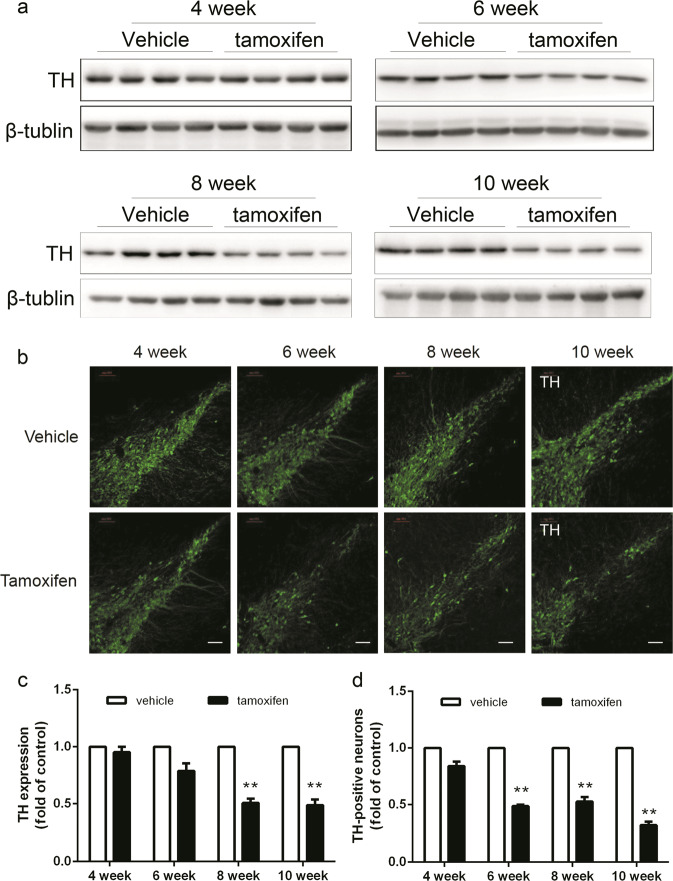
Dicer cKO mice manifested a progressive reduction on TH expression in SN. Mice were sacrificed at indicated time points after tamoxifen administration. Mice were subjected to tissue preparations for either dissecting for western assay or perfusion for immunostaining, respectively as described in Methods. a The expression of TH in SN were measured by western blot. b The immunofluorescence staining with TH in SN. Representative photomicrographs were shown at ×10 magnification (scale bars, 100 μm). c Quantitative analyses of TH expression and d counts of TH-positive neurons. Data were presented as mean ± SEM. Statistical analyses were performed by two-way ANOVA. n = 4 per group (**P < 0.01; tamoxifen vs vehicle)

