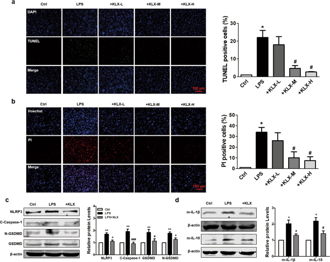
Fig. 5.
Effects of KLX on LPS-mediated NLRP3 activation and pyroptosis in cardiomyocytes. a Representative images of TUNEL staining and the quantitative analysis of TUNEL-positive cardiomyocytes from each group. The NMVCs were pre-treated with different doses of KLX for 24 h and exposed to 1 μg ·mL−1 LPS for 12 h. Scale bar = 100 μm; n = 3. b Representative images of Hoechst 33342/PI staining and quantitative analysis of PI-positive cardiomyocytes from each group. Scale bar = 100 μm; n = 3. c, d Western blot analysis of NLRP3 (n = 5), cleaved caspase-1 (n = 5), GSDMD (n = 5), N-GSDMD (n = 5), mature IL-1β (n = 4), and IL-18 (n = 5) protein levels in NMVCs treated with 10 μM KLX. The data are presented as the mean ± SEM. *P < 0.05, **P < 0.01 vs. control; #P < 0.05, ###P < 0.001 vs. LPS. a–d One-way ANOVA followed by Tukey’s post hoc analysis

