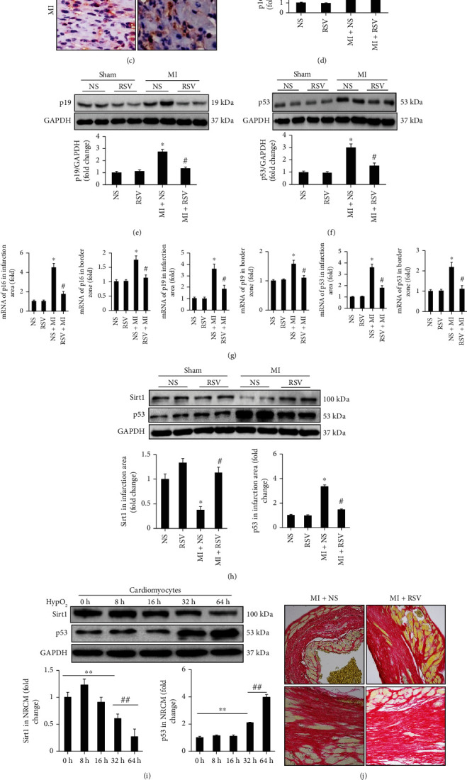
Figure 3.

RSV decreased expression of senescence markers in the mouse heart. (a–c) IHC showing the expression of senescence markers including p16, p19, and p53. (d) Representative blots and relative quantitative analysis of p16. (e) Representative blots and relative quantitative analysis of p19. (f) Representative blots and relative quantitative analysis of p53. (g) RT-PCR tested mRNA expression of p16, p19, and p53 in infarction and border zone, respectively. (h) Representative blots and relative expression level of Sirt1 and p53 in mouse heart after 3 days' MI. (i) Representative blots and relative expression level of Sirt1 and p53 in NRCMs at different time points (n = 6 in each of the NS and RSV groups, n = 6 in each of the MI and MI+RSV groups). (j) PSR staining to examine the collagen deposition. (k) mRNA expression of collagen I/III in the mouse heart tissue. (L) Western blots examined the MMP-2/MMP-9 expression. (m) Relative quantitative expression of MMP-2 and MMP-9. Mouse hearts were harvested for analysis at the 7th day after MI surgery. Data are presented as the mean ± SD. Two-way ANOVA was used for significance test. ∗p < 0.05 vs. the NS group, #p < 0.05 vs. the MI+NS group, ∗∗p < 0.05 vs. 0 h, ##p < 0.05 vs. 32 h.
