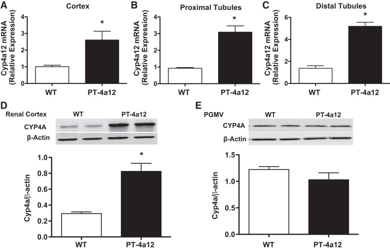
Fig. 1.
A–C: Cyp4a12 mRNA in kidney cortex (A), proximal tubules (B), and distal tubules (C) from male wild-type (WT) and PT-4a12 mice. D and E: Cyp4a protein levels in renal cortex (D) and preglomerular microvessels (PGMV; E) from male WT and PT-4a12 mice. Results are means ± SE; n = 5 mice/group. *P < 0.05 vs. WT (unpaired Student’s t test).

