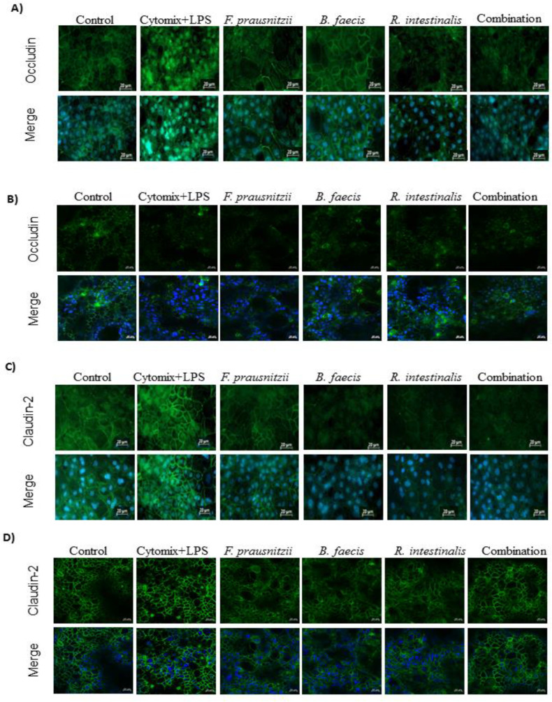
Figure 5.
Effect of F. prausnitzii, B. faecis and R. intestinalis alone and in combination on the immunofluorescence localization of tight junction proteins. Both cell line monolayers were incubated with all three bacteria alone and in combination following incubation, cell monolayers were fixed and stained with anti-occludin and claudin-2 antibodies (green) and Hoechst (blue) and imaged by confocal microscopy. Immunofluorescence staining of Occludin in Caco-2 (A) and HT29-MTX (B) cell monolayers. Immunofluorescence staining of claudin-2 in Caco-2 (C) and HT29-MTX (D) cell monolayers. Images are of 40× magnification. Bar = 20 µm.

