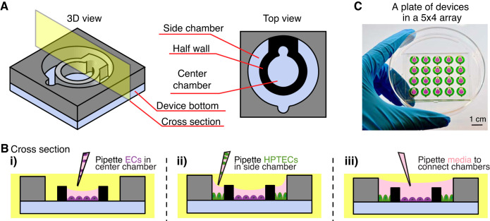
Fig. 1.
Coculture device design and operation. A: three-dimesional and top view of the device design. B: cross-sectional view of the device operation. Different cell types [endothelial cells (ECs) and human kidney proximal tubule epithelial cells (HPTECs)] are selectively seeded into the separated cell culture chambers (i and ii) and placed into paracrine signaling contact by the addition of shared media on top (iii). C: photograph of a plate of devices in the 5 × 4 array fitted inside a petri dish. Center and side chambers are loaded with purple and green dye, respectively, to visualize chamber segregation. The device loading process is shown in Supplemental Video S1.

