Fig. 2.
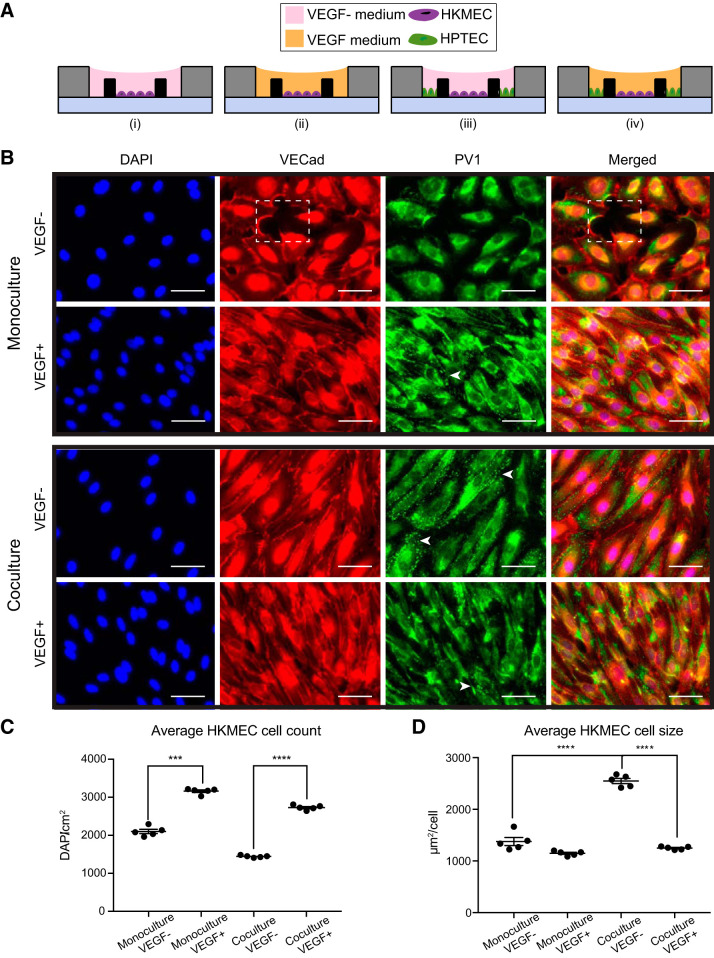
Coculture with human kidney proximal tubule epithelial cells (HPTECs) preserves human kidney peritubular microvascular endothelial cell (HKMEC) morphology in coculture. A: device cross-sectional views showing the four culture conditions of HKMECs and HPTECs: monoculture of HKMECs in media without (i) or with (ii) exogeneous VEGF and segregated coculture of HKMECs and HPTECs in media without (iii) or with (iv) exogeneous VEGF. B: immunofluorescence images of HKMECs under differential culture conditions. Blue, nuclear stain (DAPI); red, vascular-endothelial cadherin (VECad). The dotted box indicates a region without cells. Green, plasmalemma vesicle-associated protein (PV1). Arrows indicate example regions with clear PV1 structures (punctate staining). Scale bars = 50 μm. The images shown are from donor C and are representative of experiments with cells from three different human donors each with five device replicates per experiment. C: quantitative comparison of HKMEC count between “monoculture, VEGF−” and “monoculture VEGF+” conditions and “coculture, VEGF−” and “coculture, VEGF+” conditions. D: quantitative comparison of HKMEC size between “coculture, VEGF−” and “monoculture, VEGF−” conditions and “coculture, VEGF−” and “coculture, VEGF+” conditions. Error bars represent the SEs of five device replicates for a single human donor (donor C) in a single experiment. Data sets were analyzed using a one-way ANOVA test; P values are indicated for Tukey’s multiple-comparisons tests. ***P ≤ 0.001; ****P ≤ 0.0001.

