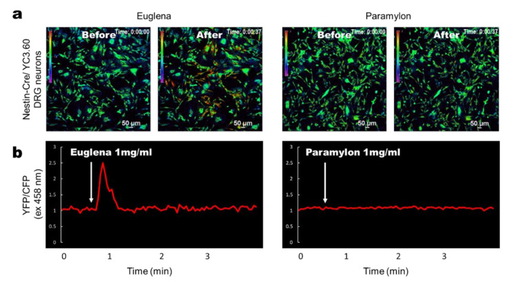
Figure 3.
Ca2+ signaling images using Euglena and Paramylon in dorsal root ganglia (DRG) neurons in vitro. (a) Representative ratiometric Ca2+ signaling images in DRG cells from YC3.60flox/Nestin-Cre mice showing YFP/CFP excitation at 458 nm. Euglena or paramylon (0.1 mL) in PBS (1 mg/mL) was added to the cell culture at the indicated time point. The color scale indicates relative Ca2+ concentration. (b) Time course of ratiometric YFP/CFP fluorescence intensity at 458 nm excitation. Results are representative of at least three independent experiments (n = 9 mice; scale bars, 50 μm).

