Fig. 3.
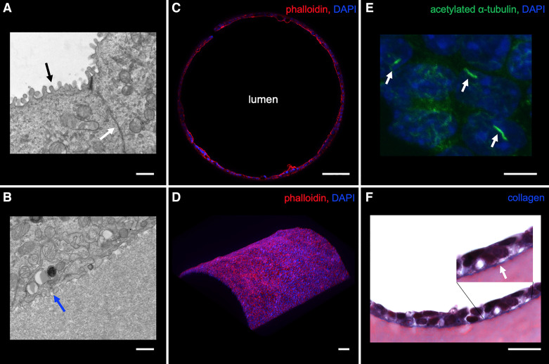
Localization of structural proteins in the 3D CCD. A and B: transmission electron microscopy of mpkCCD cells in the 3D CCD demonstrating apical microvilli (black arrow), adherent junction (white arrow), and basal infoldings (blue arrow). Scale bar = 800 nm. C: strong apical F-actin localization in mpkCCD cells grown in the 3D CCD. Scale bar = 100 μm. D: 3D reconstruction revealing a curved epithelial monolayer as identified by F-actin staining. Scale bar = 100 μm. E: cells grown in the 3D CCD had immunodetectable cilia. Scale bar = 10 μm. F: cells deposit collagen along the basal surface onto the extracellular matrix (ECM) as identified by linear blue staining with Masson’s trichrome and visualized by light microscopy. Scale bar = 20 μm.

