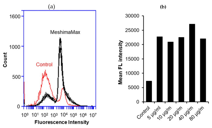
Figure 7.
Internalization of fluorescence nano-beads by macrophages under the treatment of MeshimaMax. The flow cytometry results are shown in (a) where MeshimaMax treatment induced a remarkable shift to a higher fluorescence intensity than untreated controls indicating that more nano-beads were taken up by macrophages. The mean fluorescence intensities of macrophages treated by different concentrations of MeshimaMax are shown in (b). See text for details.

