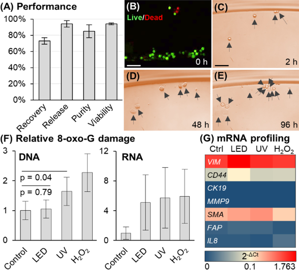
Figure 2.
(A) Performance of sinusoidal microfluidic device using PC linker for anti-EpCAM enrichment of SKBR3 cells spiked into whole blood (N = 3). (B) LED release had no effect on viability, and (C–E) released cells in culture for 2–96 h (Scale bars = 100 μm). (F) DNA/RNA oxidative damage (N = 3) assessed for 2 min LED exposure, equivalent UV dose (18.5 J), and 300 μM H2O2 (30 min) of Hs578T cells. DNA and RNA damage is normalized to control. (32.2 pg 8-oxo-G per 400 ng DNA and 7.15 pg 8-oxo-G per 400 ng RNA). The DNA-derived ELISA 8-oxo-G calibration curve could not quantify RNA damage absolutely. (G) mRNA profiling by RT-qPCR (N = 3) of Hs578T cells following no irradiation, LED or UV light exposure, or H2O2 treatment.

