Fig. 4.
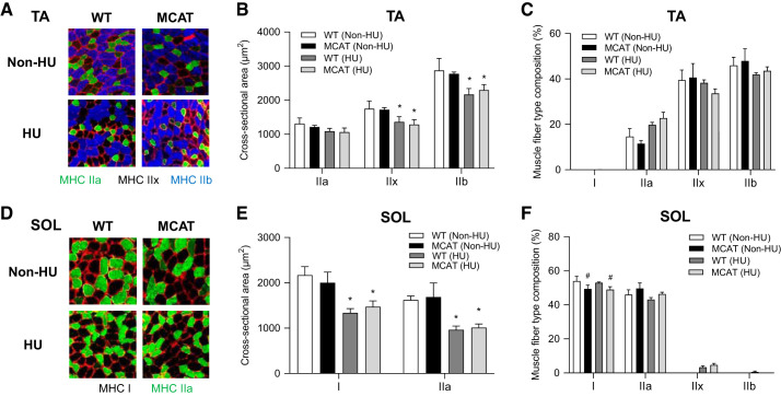
Mitochondrial-targeted catalase (MCAT) has no effect on muscle fiber cross-sectional area. A: representative images of myosin heavy chain (MHC) immunofluorescence for tibialis anterior (TA) muscles in wild-type (WT) and MCAT mice with or without HU. B: muscle fiber cross-sectional area by fiber type for TA muscles. C: fiber-type composition for TA muscles (WT non-HU, n = 4; MCAT non-HU, n = 4; WT HU, n = 6; MCAT HU, n = 6). D: representative images of MHC immunofluorescence for soleus (SOL) muscles. E: muscle fiber cross-sectional area by fiber type for SOL muscles. F: fiber type composition for SOL muscles (WT non-HU, n = 3; MCAT non-HU, n = 3; WT HU, n = 7; MCAT HU, n = 8). *Main effect of HU (P < 0.05); #main effect of genotype (P < 0.05). Values are means ± SE.

