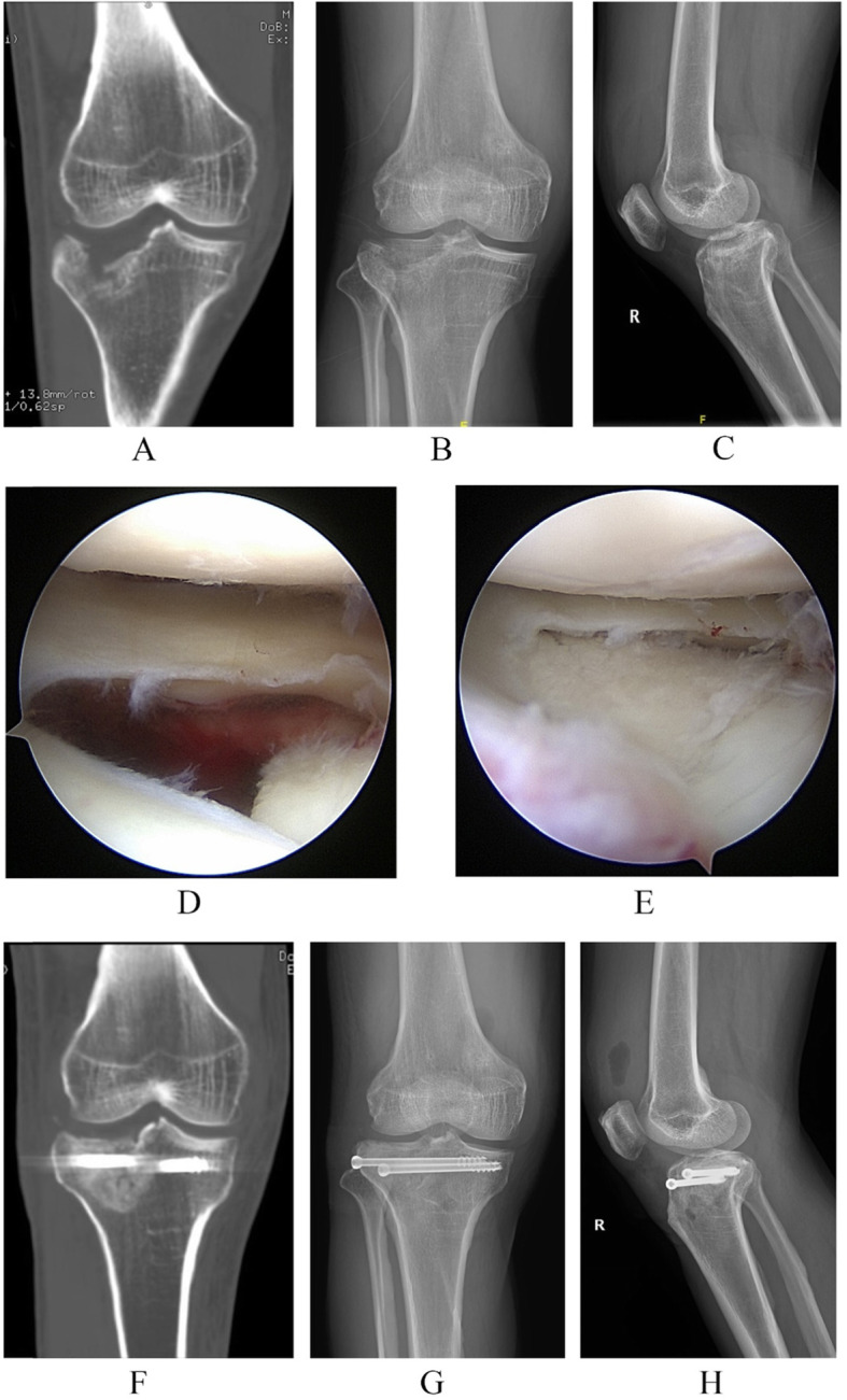
Fig. 3.

A 44-year-old male patient who was diagnosed with the lateral inclination type of posterolateral tibial plateau fracture with LI patterns. a The coronal image of three-dimensional CT scans shows the lateral inclination of the tibial fracture fragment. b The anteroposterior (AP) plain film of the injured knee. c The lateral plain film of the injured knee. d The tibial plateau fracture visualized with the assistance of arthroscopy. e After restoration of the fracture fragments with the surveillance of arthroscopy, the step-off was eliminated. f The coronal image of three-dimensional CT scans shows the restoration and the fixation. g The post-operation AP film of; H, the post-operation lateral plain film
