Fig. 2.
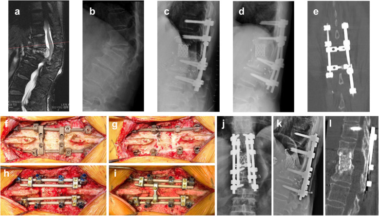
Case 18. A 66-year-old woman with breast cancer metastasis at L1. a Preoperative sagittal T2 magnetic resonance image revealing tumor involvement at L1. b Lateral radiograph revealing pathological fracture at L1. c Lateral radiograph after total en bloc spondylectomy. d Radiographs at 18 months after surgery revealing increased local kyphosis angle between T12 and L2. e Coronal computed tomography (CT) scan revealing breakage of the left rod. f Posterior instrumentation is exposed using the previous midline incision. g The left rod is broken at the area proximal to the L2 pedicle screw. h Loosening pedicle screw at the left T11 level is replaced, and four cobalt chromium rods are inserted. i Autologous bone graft is placed on adjacent T12 and L2 lamina and scar tissue of the resected L1 lamina area. j, k Radiograph after revision surgery. l Sagittal CT scan at 42 months after revision surgery revealing bone fusion within the cage and at the posterior bone graft

