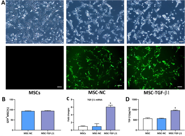
Fig. 1.
Measurement of TGF-β1 in genetically modified MSCs. a Green fluorescent protein was expressed in lentivirus-infected MSCs. Light microscopy (top) and fluorescence microscopy (bottom) images. Scale bar = 100 μm. b Quantitative analysis of lentivirus transduction. c TGF-β1 mRNA expression in MSCs, MSC-NC, and MSC-TGF-β1. d The expression of TGF-β1 in 2 × 10⁶ MSCs, MSC-NC, or MSC-TGF-β1 in 200 mL of culture supernatant, as measured by ELISA (n = 3; &p < 0.05 vs. MSC-NC). The means ± SD of three experiments is shown. ELISA, enzyme-linked immunosorbent assay; MSCs, mesenchymal stem cells; MSC-NC, mesenchymal stem cell carrying GFP; MSC-TGF-β1, TGF-β1 overexpressing MSC

