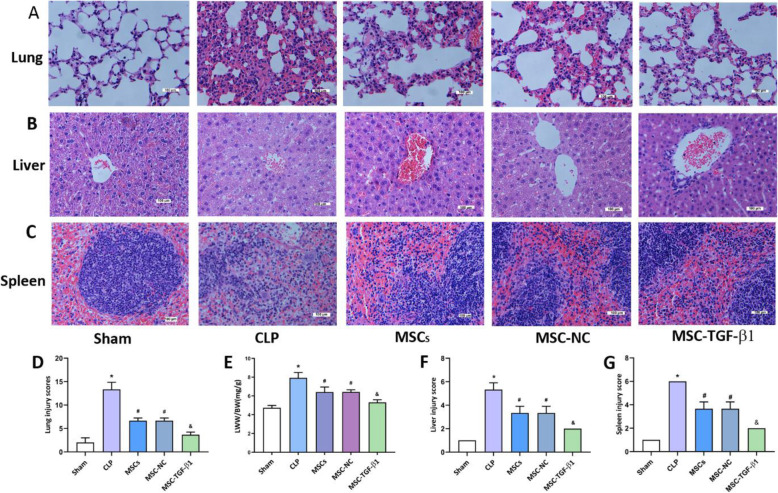
Fig. 2.
The effect of MSC-TGF-β1 on organ injury in CLP-induced septic mice. a–c Histopathological images of lung, liver, and spleen tissues (H&E staining, × 400). Scale bar = 100 μm. d The injury scores of the lung tissue. e Pulmonary capillary permeability was measured by the LWW/BW ratio. f, g The injury scores of the liver and spleen (n = 3; *p < 0.05 vs. sham group; #p < 0.05 vs. CLP group; &p < 0.05 vs. MSC-NC group). CLP, cecal ligation and puncture; MSCs, mesenchymal stem cells; MSC-NC, mesenchymal stem cell carrying GFP; MSC-TGF-β1, TGF-β1 overexpressing MSC

