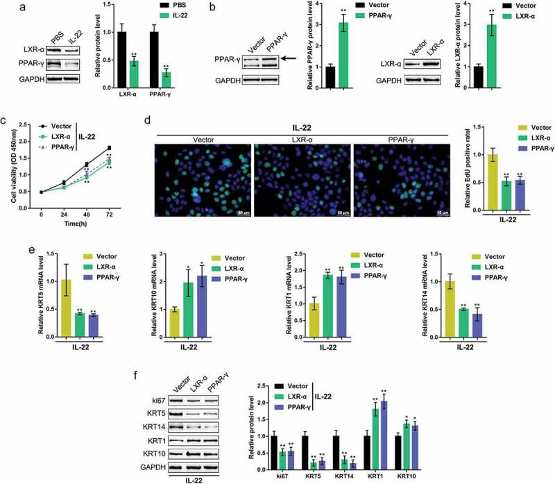
Figure 2.

Effects of LXR-α and PPAR-γ on keratinocytes HaCaT cells were treated with IL-22 to generate a psoriasis-like dermatitis model in vitro and examined for (a) the protein levels of LXR-α and PPAR-γ using immunoblotting. (b) an LXR-α or PPAR-γ-overexpressing vector was transfected into HaCaT cells, and the transfection efficiency was verified by western blot (n = 3). Next, HaCaT cells were transfected with the LXR-α or PPAR-γ-overexpressing vector, treated with IL-22, and examined for (c) cell viability using the MTT assay (n = 5); (d) DNA synthesis capacity by the EdU assay (n = 3); (e) the mRNA expression of KRT5, KRT10, KRT1, and KRT14 using real-time PCR (n = 5); and (f) the protein levels of ki67, KRT1, KRT10, KRT5, and KRT14 using immunoblotting (n = 3). *P < 0.05, **P < 0.01.
