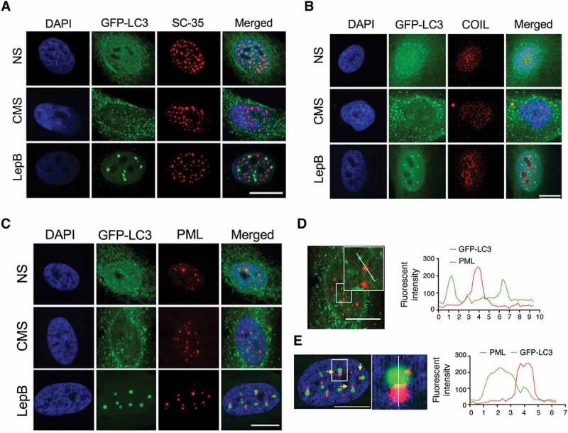
Figure 4.

LC3 interacts with PML nuclear bodies in LepB-treated cells, but not with CMS-induced ones. hTM cells were transduced with AdGFP-LC3 (5 pfu/cell) and either exposed to CMS (8% elongation) or LepB (20 nM) treatment for 24 h. Cells were fixed and immunostained with specific antibodies against nuclear speckels (A), Cajal bodies (B) and PML bodies (C). Fluorescent intensity of GFP-LC3 signal (green) and PML (red) in CMS (D) and LepB-treated cells (E) were quantified with Fiji software. DAPI was used to stain nuclei. Scale bars: 20 μm.
