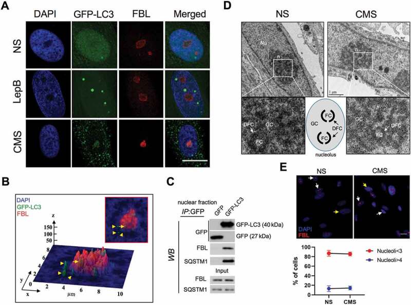
Figure 5.

Nuclear LC3 associates with the nucleolus with CMS. (A) Representative immunostaining of the nucleolar marker FBL in AdGFP-LC3-transduced cells exposed for 24 h to CMS (8% elongation) or LepB (20 nM) treatment. DAPI was used to stain nuclei. Scale bars: 20 μm. (B) Interactive 3D surface plot analysis visualizing the interaction between GFP-LC3 and the nucleolus. (C) Co-IP analysis of GFP-LC3 with FBL and SQSTM1 in purified nuclear fractions from hTM cells transduced with AdGFP or AdGFP-LC3. IP was performed using GFP-Trap; co-immunoprecipitated proteins were detected by WB, 5 µg of protein were loaded for input control. (D) Representative EM images depicting nucleolar structure and components in NS and cells under CMS. FC: Fibrillar Center; DFC: dense fibrillar component; GC: granular component. (E) Quantification of nucleolar number in NS and cells under CMS. Nucleoli were detected by immunostaining with FBL antibody (red). Y axis represents percentage of cells containing nucleoi number>4 per nucleus in total cells counted. Data are shown as the mean ± S.D. (n = 375 and 309 in CNT and CMS, respectively), two-tailed unpaired Student’s t-test.
