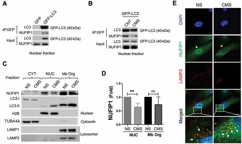
Figure 7.

Nuclear LC3 interacts with the ribophagy receptor NUFIP1 and promotes its nucleocytoplasmic distribution in hTM cells with CMS. Co-IP analysis in nuclear fraction of control cells transduced with (A) AdGFP or AdGFP-LC3 or (B) AdGFP-LC3 expressing cells with or without CMS. IP was performed using GFP-Trap; co-immunoprecipitated proteins were detected by WB, 5 µg of protein were loaded for input control. (C) WB blot analysis of NUFIP1 and LC3 in fractionated cytosolic, nuclear and membrane-bound organelle enriched fractions from NS and hTM cells under CMS. TUBA4A and H2B are used as a loading controls for cytosolic and nuclear fraction, respectively. LAMP1 and LAMP2 were used as markers for membrane-bound organelle enriched fractions containing lysosomes. (D) NUFIP1 band intensity was normalized and fold expression calculated. Data are shown as the mean ± S.D. (n = 3), **, p < 0.01, two-tailed unpaired Student’s t-test. ns: not significant. (E) Representative immunostaining of NUFIP1 (green) and LAMP2 (red) in NS and hTM cells subjected to CMS (8% elongation, 24 h). Yellow arrowheads indicate nuclear NUFIP1 staining; white arrows indicate co-localization of NUFIP1 and LAMP2.
