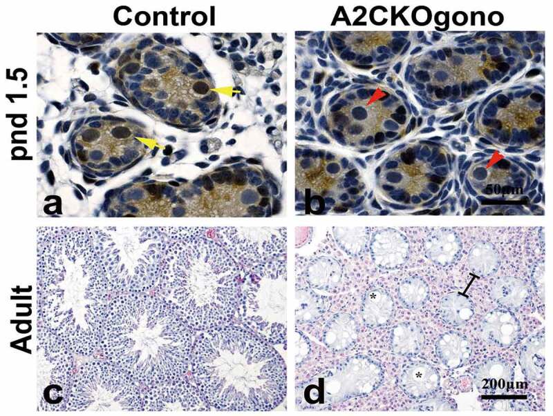
Figure 2.

Loss of cyclin A2 expression in gonocytes results in severely abnormal spermatogenesis and few if any germ cells in the adult testis. (a,b) Testicular sections from control and A2CKOgono at pnd 1.5 were stained with anti-cyclin A2 antibody using immunohistochemistry. The yellow arrows indicate cyclin A2 positive germ cells and red arrows indicate the cyclin A2 negative germ cells. Bar indicates 50 μm. (c,d) H&E staining in adult mouse testes from control (c) and A2CKOgono (d). The black lines indicate the hypertrophy of the interstitial tissue. Bar indicates 200 μm. The asterisks indicate the vacuolation of the tubules.
