
Figure 5.
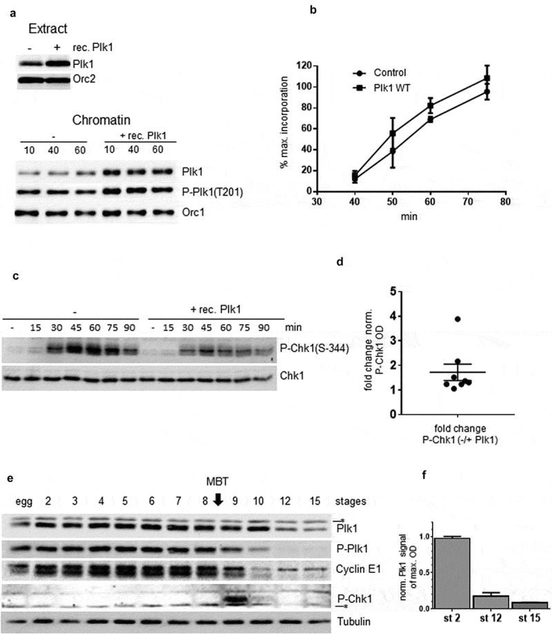
Accelerating effect of Plk1 overexpression on DNA replication and changes in Plk1 abundance during early Xenopus development A Western blot analysis of Plk1 in egg extract (upper) or in chromatin fractions (lower) in control or after Plk1 addition. B Sperm nuclei (2000 n/µl) were incubated in the presence of α32P-dCTP and recombinant Plk1 and reactions were stopped at indicated time, then DNA was purified and analyzed by alkaline gel electrophoresis, mean of percent of maximal incorporation from three independent experiments. C Western blot analysis of P-Chk1 and Chk1 at different time (min) during S phase with (+) or without (-) addition of recombinant Plk1 addition in whole extract. D quantification of 8 different P-Chk1 optical densities of western blots from two independent S phase kinetics experiments normalized with Chk1, points indicating fold change of P-Chk1 from control over Plk1 overexpression samples, with mean (1.722) and standard error of mean 0.33 (SEM). E Xenopus embryos were harvested after in vitro fertilization at indicated stages and whole embryo lysates were analyzed by western blot with Plk1, P-Plk1(T201), cyclin E1, P-Chk1(S344) and tubulin antibodies. F Quantification of western blot, optical densities (OD) of Plk1 normalized to tubulin, at stage 2, 12 and 15 from three different in vitro fertilizations, different egg batches from three female frogs.