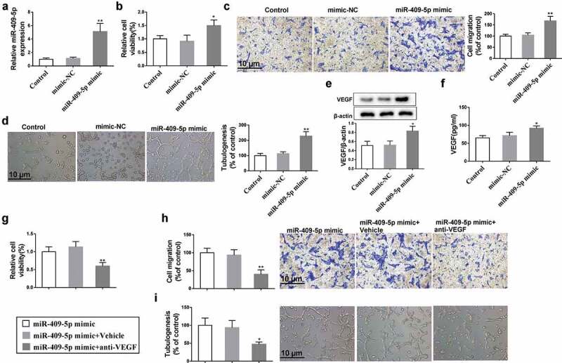
Figure 3.

Effect of overexpressing miR-409-5p on neovascularization. mRMECs were transfected with miR-409-5p mimic or the negative control (mimic-NC) for 48 h. (a) The expression of miR-409-5p was detected using qRT-PCR. (b) Cell viability was detected using cell counting kit-8 (CCK-8) assay. (c) Cell migration was detected using transwell migration assay. (d) Tube formation was observed at 6 h under a microscope. (e) The expression of VEGF in mRMECs was detected using Western blot analysis. (f) The expression of VEGF in supernatants of culturing medium was detected using ELISA. mRMECs were transfected with miR-409-5p mimic for 48 h, followed by the treatment of VEGF antibody (100 μg/mL) or the control Vehicle (same volume of PBS) for 24 h. Cell viability (g), cell migration (h), and tube formation (i) were detected. *p < 0.05, **p < 0.01 vs mimic-NC or Vehicle. Three samples in each bar graph. Scale bar = 10 μm. Magnification = 200 ×.
