Figure 1.

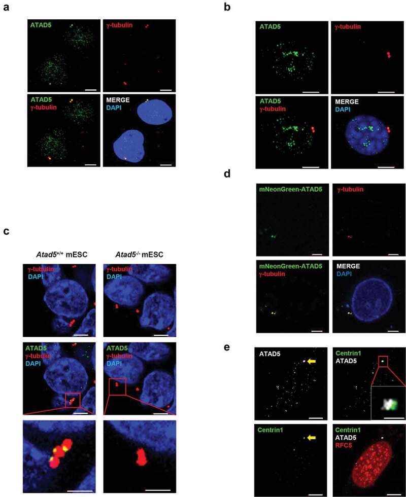
ATAD5 is localized at the centrosomes in an RFC independent manner.
(a) U2OS cells and (b) NIH3T3 cells were fixed at asynchronous condition for immunostaining. Bar, 5 μm. (c) The wild type (Atad5+/+) mouse embryonic stem cells (mESCs) and Atad5 knock out (Atad5−/-) mESCs were fixed for immunostaining. Bar, 5 μm. The inset shows high-magnification image of the centrosome. Bar, 2 μm. (d) U2OS cells were transfected with a DNA vector expressing mNeonGreen-fused ATAD5 N-terminal fragment (1–693 amino acids). After 48 h of transfection, cells were fixed for immunostaining as indicated. Bar, 5 μm. (e) HeLa cells stably expressing GFP-tagged Centrin1 (HeLa_Centrin1-GFP) were fixed at asynchronous condition for immunostaining. The arrow indicates a centrosome. Bar, 5 μm. The inset shows high-magnification image. Bar, 1 μm.
