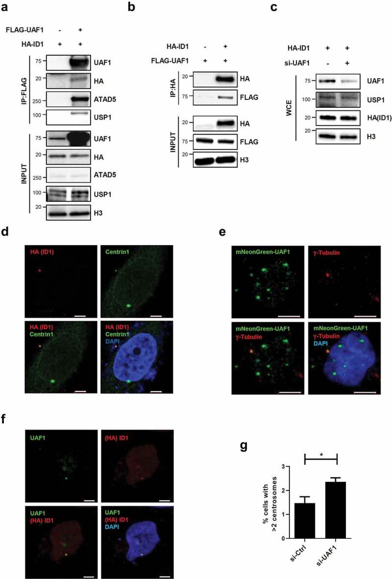
Figure 5.

UAF1 and ID1 interact, and co-localize at the centrosome.
(a, b) HEK293 T cells were transfected with 3xFLAG-tagged UAF1 cDNA (FLAG-UAF1) and HA-tagged ID1 cDNA (HA-ID1). (a, b) After 48 h, whole cell extracts were prepared for immunoprecipitation (IP) with an anti-FLAG antibody (A) and an anti-HA antibody (b). Immunoprecipitates were eluted, resolved by SDS-PAGE, and immunoblotted using the indicated antibodies. (c) HEK293 T cells were transfected with a combination of UAF1 siRNA and HA-ID1 cDNA. After 48 h of transfection, whole cell extracts (WCE) were subjected for immunoblotting. (d-f) After 48 h of transfection, cells were fixed for immunostaining as indicated. HeLa_Centrin1-GFP cells (d) and HeLa cells (f) were transfected with HA-ID1 cDNA. (e) HeLa cells were transfected with mNeonGreen-fused UAF1 cDNA. Bar, 5 μm. (g) HeLa_Centrin1-GFP cells were transfected with UAF1 siRNA for 48 h and fixed. The percentage of cells with more than two centrosomes was displayed. Three independent experiments were performed. Values represent mean ± SEM. Statistics: paired t-test. *, p ≤ 0.05.
