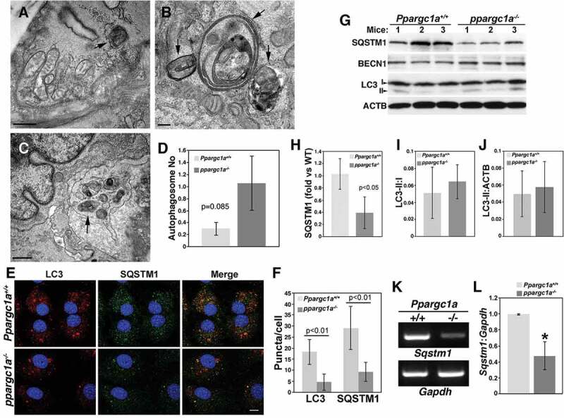
Figure 3.

Abnormal autophagosomes and reduced SQSTM1 expression in the aorta of ppargc1a−/- mice. Aortas of Ppargc1a+/+ (A) and ppargc1a−/- mice (B and C) were analyzed by TEM. Arrows indicate autophagosomes. Bar: 0.5 μm in A and C, and 0.2 μm in B. Graph presenting the overall average of autophagosomes in both genotypes is shown in D. Ppargc1a+/+ and ppargc1a−/- VSMCs were fixed and incubated with mouse LC3 and rabbit SQSTM1 antibodies (E). Images in E were acquired using a confocal microscope for the quantification of LC3 and SQSTM1 puncta. Bar: 10 μm. (F). 34 cells and 30 cells were analyzed for LC3 in WT and knockout, respectively, while 23 and 24 cells were analyzed for SQSTM1 puncta in WT and knockout, respectively. Aortas of 6-month-old Ppargc1a+/+ and ppargc1a−/- male mice were lysed and processed for western blot analysis (G). SQSTM1, LC3-I and LC3-II levels were adjusted by ACTB and expressed as fold change, compared with WT (H-J). mRNA was isolated from Ppargc1a+/+ and ppargc1a−/- VSMCs and used to measure Sqstm1 and Gapdh mRNA levels (K and L). 3 independent mRNA preparations per genotype. * denotes p < 0.01 in L.
