
Figure 1.
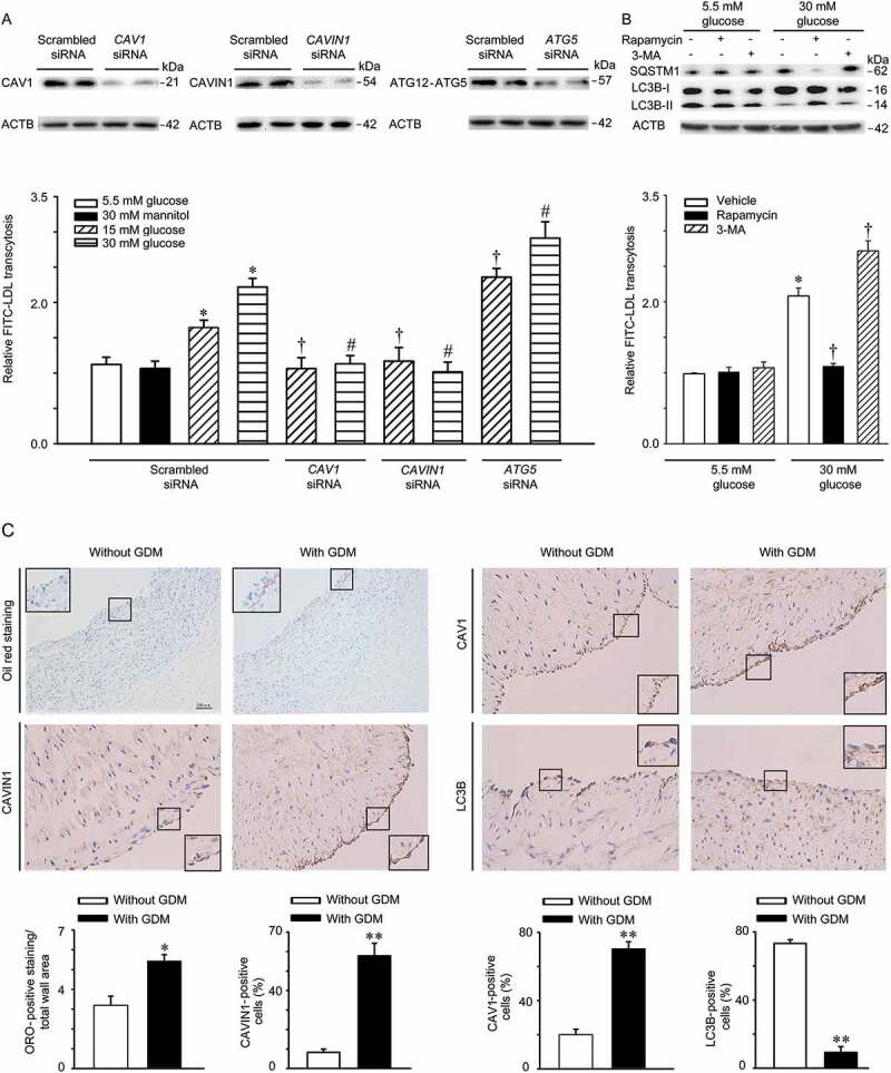
High glucose stimulates LDL transcytosis. (A) HUVECs were transfected with scrambled siRNA (20 nM), CAV1 siRNA (20 nM), CAVIN1 siRNA (20 nM), or ATG5 siRNA (20 nM) for 48 h, followed by treatment with glucose (5.5 mM, 15 mM or 30 mM) or mannitol (5.5 mM glucose+24.5 mM mannitol) for 24 h and FITC-LDL treatment for 3 h. LDL transcytosis was evaluated. (B) HUVECs were treated with rapamycin (5 nM, 24 h) or 3-MA (5 mM, 4 h) with different concentrations of glucose (5.5 mM, 30 mM, 24 h), after which LDL transcytosis was evaluated. * p < 0.05 versus scrambled siRNA +5.5 mM glucose or 5.5 mM glucose alone; † p < 0.05 versus scrambled siRNA+15 mM glucose or 30 mM glucose alone, # p < 0.05 versus scrambled siRNA+30 mM glucose, (n = 4). (C) Human umbilical cords were stained with Oil Red O (ORO), CAV1, CAVIN1, and LC3B, respectively. Quantitative summary of the percentage of area Oil Red or CAV1-CAVIN1-LC3B-positive cells in human umbilical cords venous. Scale bars: 100 μm. * p < 0.05, ** p < 0.01, versus without GDM.