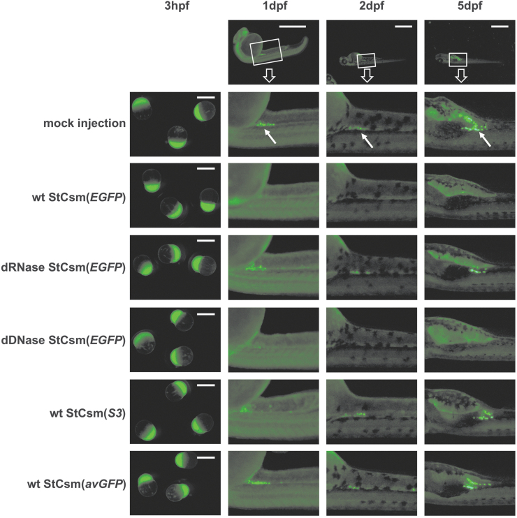
FIG. 2.
Microscopy of StCsm mediated EGFP knockdown in Tg(ddx4:ddx4-EGFP) fish. Fluorescence from Tg(ddx4:ddx4-EGFP) fish after injection with wildtype (wt) or mutant StCsm complexes are shown. The arrows indicate the location of primordial germ cells. Injection was done at the 1-cell embryo stage, observations were at 3 hpf, 1 dpf, 2 dpf, and 5 dpf. The scale bar represents 1 mm. Boxes in the top row show the region of the embryo magnified in panels below. Arrows point to germ cells.

