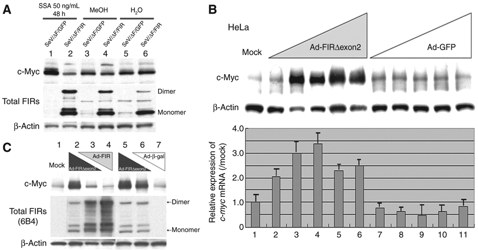
Figure 4.

Ad–FIR suppressed SSA-activated c-Myc, whereas Ad–FIRΔexon2 activated c-myc transcription and led to c-Myc overexpression. A, c-Myc activation in 50 ng/mL SSA treatment for 48 hours was suppressed by enforced SeV/ΔF/FIR expression (lane 2). The effect of SeV/ΔF/FIR (27) was examined for whether the increase in c-Myc from either the SAP155 siRNA or SSA treatment is impaired by FIR. SeV/ΔF/FIR suppressed SSA-induced c-Myc activation (compare lane 2 with lane 1), but not basal c-Myc expression (compare lanes 4 and 6 with lanes 3 and 5, respectively). B, HeLa cells were treated with Ad–FIRΔexon2 for 48 hours, and whole cell proteins and total RNAs were extracted. Western blot analysis for c-Myc protein expression and qRT-PCR for c-myc mRNA were carried out. Ad–FIRΔexon2 activated c-Myc protein and c-myc mRNA in HeLa cells. Ad–GFP was used as a control vector. Mock shows that HeLa crude extract proteins are not subject to adenovirus vector treatment. Lanes 2, 7: 0.1 MOI; lanes 3, 8: 0.5 MOI; lanes 4, 9: 1 MOI; lanes 5, 10: 5 MOI; lanes 6, 11: 10 MOI of Ad–FIRΔexon2 or Ad–GFP, respectively. Ad–FIRΔexon2 apparently increased c-Myc more than 20 times over mock expression, whereas Ad–GFP did not affect c-Myc. The increase of c-myc mRNA was much less, 2 to 3 times that of c-Myc protein elevation by Ad–FIRΔexon2. C, FIR and Ad–FIRΔexon2 antagonized against c-Myc expression (compare lanes 3 and 6). Lane 1 (Mock): no adenovirus vector; Lane 2: 10 MOI of Ad–FIRΔexon2; Lane 3: 5 MOI of Ad–FIRΔexon2 and 5 MOI of Ad–FIR; Lane 4: 10 MOI of Ad–FIR; Lane 5: 10 MOI of Ad–FIRΔexon2 (same as lane 2); Lane 6: 5 MOI of Ad–FIRΔexon2 and 5 MOI of Ad–β-gal; Lane 7: 10 MOI of Ad–β-gal. Notably, c-myc mRNA was not significantly activated by Ad–FIRΔexon2 (data not shown). MOI, multiplicity of infection.
