
Figure 2. Proteins that contribute to invadopodium structure and function.
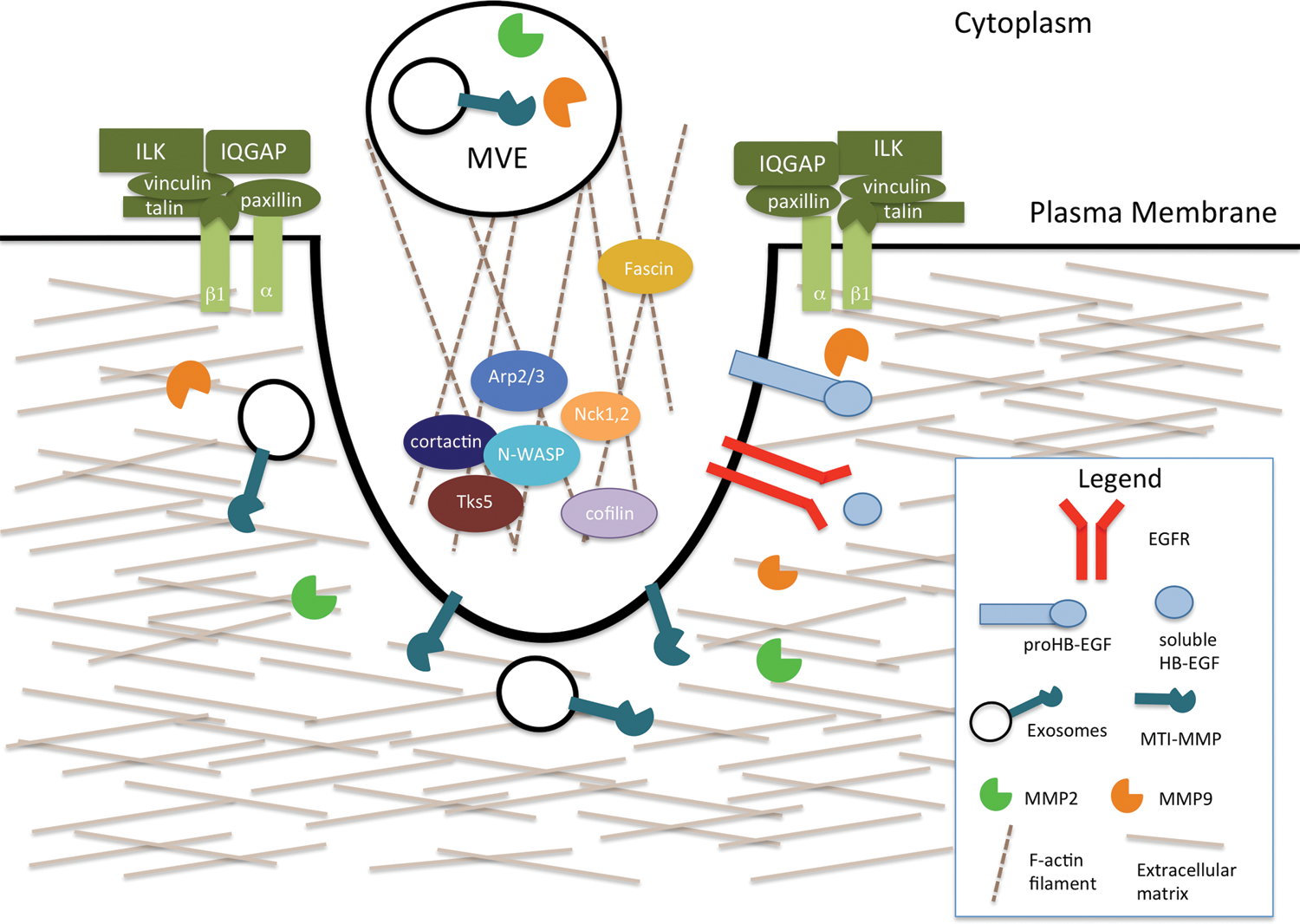
Invadopodia are actin-rich structures that specialize in mediating the degradation of the ECM. Their formation can be induced by EGFR signaling. The key components of the core structure are cortactin, Tks5, N-WASP, Arp2/3, Nck-1 and −2, and cofilin. Adhesion rings, made up of adhesion proteins such as integrins and ILKs, are important for the formation and stabilization of invadopodia. Proteases, such as MMP2 and MMP9, are secreted at invadopodia. MT1-MMP can be transported by exosomes, which are secreted from multi-vesicular endosomes.