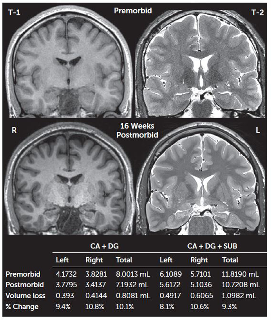
FIGURE 2. Pre- and postmorbid MRI of the hippocampi in a patient with opioid-related acute amnestic syndromea.

a CA=cornu ammonis, DG=dentate gyrus, SUB=subiculum. Volumetric units are in milliliters (mL). The total intracranial and total gray matter volumes premorbid versus postmorbid differed by approximately 1% (1745.5589 mL, 1764.6615 mL, 837.5098 mL, and 848.7063 mL, respectively).
