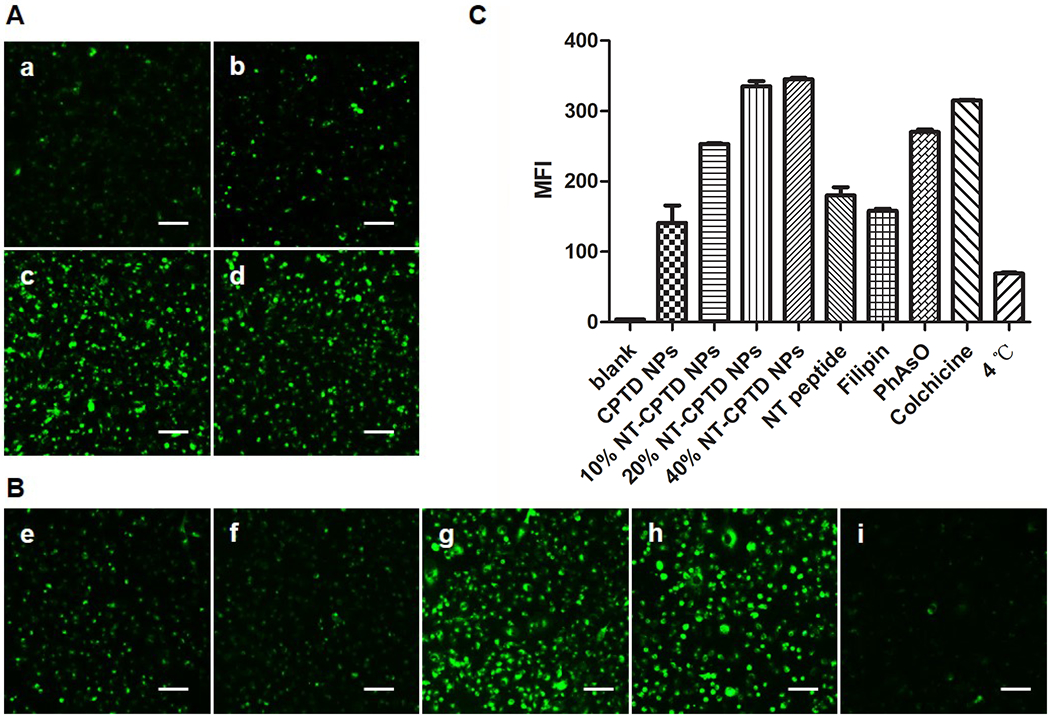
Figure 4.

(A) Cellular uptake of (a) CPTD NPs and NT-CPTD NPs with different NT-PEG5000-CPTD ratios (b) 10, (c) 20, and (d) 40 wt% in MDA-MB-231 cells after 30 min incubation. (B) Possible endocytosis pathway of NT-CPTD NPs study. Cells were blocked by different inhibitors (e) 50 μM NT peptide (f) 1 μg/mL filipin (g) 0.3 μg/mL PhAsO (h) 1 μg/mL Colchicine (i) Cells were incubated at 4 °C. Scale bars represent 100 μm. (C) Quantitative results of (A) and (B) analyzed from Flow cytometry analysis.
