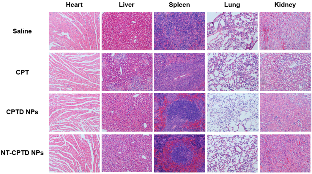
Figure 8.

Pathological examination of organs resected from MDA-MB-231 tumor-bearing mice treated with different CPT formulations. Saline group was served as control. Nuclei were stained by hematoxylin (blue), and extracellular matrix and cytoplasm were doped by eosin (red).
