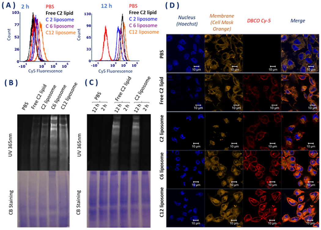
Figure 2.

(A) Flow cytometry analysis of the cellular internalization of the liposomes after 2 and 12 h incubation with the liposome and free C2 lipid (AAM). (B) SDS-PAGE analysis of azide groups in glycoproteins of MDA-MB-231 cells in vitro after 2 and 12 h incubation with the liposomes compared to free C2 lipid (AAM). The gel was detected under 365 nm UV directly after DBCO-Cy5 incubation for another 1 h. The Coomassie stain showed the total amount of protein. (C) SDS-PAGE analysis of azide groups in glycoprotein of MDA-MB-231 cells in vitro after 2 and 12 h incubation with free C2 lipid and C2 liposome. The gel was detected under 365 nm UV directly after DBCO-Cy5 incubation for another 1 h. The Coomassie stain showed the total amount of protein. (D) Confocal microscopy images of MDA-MB-231 cells incubated with 10 μM of Ac3ManNAzOH-lipid-loaded liposomes and free AAM for 12 h at 37°C, followed by labeling with DBCO-Cy5 for another 1 h. Cells treated with PBS and labeled with DBCO-Cy5 were used as the control. The cell nucleus was stained with Hoechst and the membrane was stained with cell mask orange.
