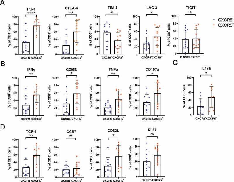
Figure 4.

Phenotyping intratumoral CXCR5+CD8+ T cells in MIBC patients.
(a) Comparison of immune checkpoints (PD-1, CTLA-4, TIM-3, LAG-3 and TIGIT) expression on CXCR5+CD8+ T cell vs. CXCR5− CD8+ T cell in MIBC. (b) Comparison of effector cytokines (IFN-γ, GZMB, TNF-α and CD107a) expression on CXCR5+CD8+ T cell vs. CXCR5− CD8+ T cell in MIBC. (c) Comparison of IL-17A expression on CXCR5+CD8+ T cell vs. CXCR5− CD8+ T cell in MIBC. (d) Comparison of TCF-1, CCR7, CD62L and Ki-67 expression on CXCR5+CD8+ T cell vs. CXCR5− CD8+ T cell in MIBC. Data was analyzed by Pearson correlation test and Student t test. *P < .05, **P < .01, ***P< .001, ****P < .0001.
