Figure 1.

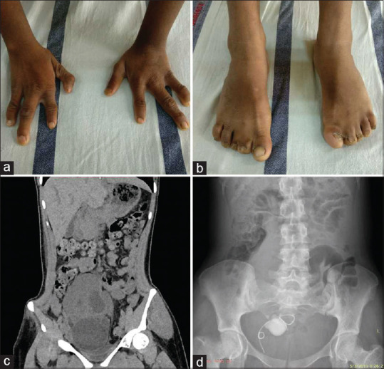
(a) Bilateral hands showing oligodactyly. (b) Bilateral foot showing syndactyly. (c) CT abdomen showing large solitary ectopic pelvic kidney with hydronephrosis. (d) Plain X-ray KUB showing DJ stent in situ and renal calculi

(a) Bilateral hands showing oligodactyly. (b) Bilateral foot showing syndactyly. (c) CT abdomen showing large solitary ectopic pelvic kidney with hydronephrosis. (d) Plain X-ray KUB showing DJ stent in situ and renal calculi