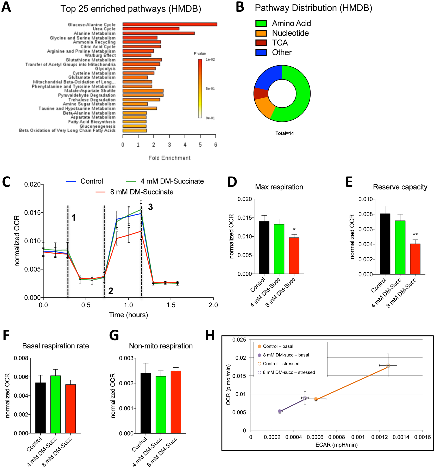
Figure 5: Succinate elevation impairs mitochondria function in myoblast cells.

(A) A bar chart depicting pathway enrichment analyses (sorted by p-value) using HMDB IDs. Shown are the top 25 enriched processes. All data are derived from Metaboanalyst processing of non-targeted analysis of C2C12 cells treated with control or 8 mM DM-succinate media for 48 hours. n=3 control and n=3 8 mM DM-succinate. (B) A circular pie chart depicting the relative distribution of differentially regulated metabolic pathways using HMDB IDs during function explorer analysis. (C) Line graphs showing oxygen consumption rates (OCR), normalized to cell count, of C2C12 myoblasts treated sequentially with oligomycin (dashed line 1), FCCP (dashed line 2), and Antimycin A/Rotenone (dashed line 3) in the presence or absence of 4 mM or 8 mM DM-succinate. (D-G) Bar graphs displaying basal respiration rates (F), maximal respiration (G), reserve capacity (H), and non-mitochondrial respiration (I). n=3; *p<0.05, **p<0.01 by unpaired student’s t-test. (H) Line graphs depicting OCR and ECAR (extracellular acidification rate) in response to mitochondria stress (Energy phenotype assay; Seahorse) in C2C12 control or DM-succinate treated myoblasts. Filled circles are basal conditions, open circles are stressed conditions. n=3 control and n=3 8 mM succinate.
