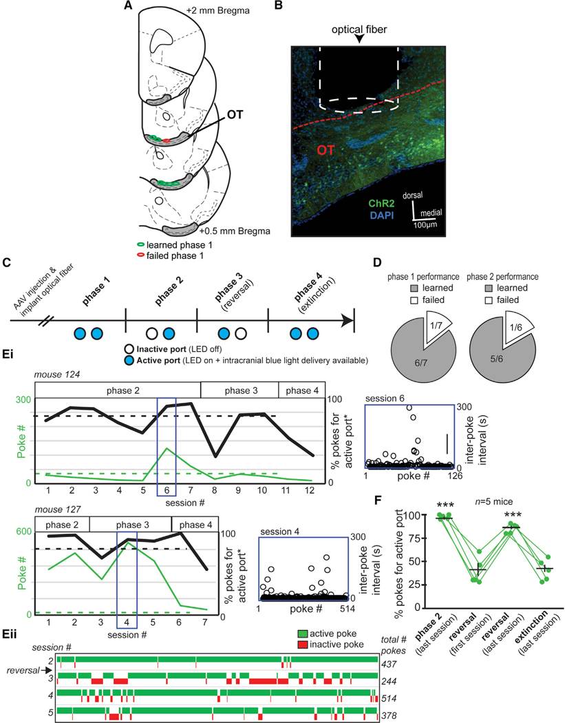
Figure 6. OT Neurons Expressing the D1 Receptor Promote Behavioral Engagement.
(A) Optical fiber implant locations from mice contributing optical-intracranial self-stimulation (opto-ICSS) data, segregated based on those mice that learned versus failed to acquire phase 1 of the task.
(B) Representative image of an optical fiber positioned within/immediately above the OT of a D1-Cre mouse that was previously injected with AAV.ChR2. Red dashed line indicates the dorsal border of the OT. Scale bar, 100 μm.
(C) Opto-ICSS task schematic wherein mice were required to nose poke in exchange for blue-light-mediated optogenetic excitation of OT D1 neurons. At least 2 weeks following injection with either a floxed AAV encoding ChR2 and a reporter fluorophore (AAV.ChR2) or a floxed AAV solely encoding a reporter fluorophore (AAV), mice were shaped on the opto-ICSS task. Please see Results for a description of the four task phases.
(D) Pie chart indicating that all but one AAV.ChR2 mouse reached criterion performance on phase 1 of the task.
(E) In (i), performance of two example mice during phases 2–4 of the opto-ICSS task is indicated. The mice displayed diversity in their number of pokes into the active port and in the percentage of pokes displayed for the active (blue-light-emitting) versus inactive ports. Both of these mice reached criterion on phase 2 and phase 3 wherein they had to learn to redirect their poking for a new active port location (reversal learning). Indicated also is the behavior of these two mice during extinction, wherein the port lights were on (viz., both ports were visually “active”) but no optogenetic stimulation was available regardless of poking. Asterisks indicated data plotted as “active port” referring to the previously active port in phase 3. Blue insets illustrate each animal’s inter-poke intervals (open circles) throughout the indicated behavioral session. (ii) A win-loss plot for mouse 127 from sessions 2–5.
(F) Quantification of opto-ICSS data indicating that ChR2-mediated stimulation of OT D1 neurons promotes task engagement. The first phase of reversal learning resulted in a significant reduction in the percentage of pokes for the new active port, which, with experience, was restored on the last session of the reversal phase and again reduced during subsequent extinction testing. Data points indicate individual mice. Green data points indicate individual mice, with the population averages (±SEM) overlaid. ***p < 0.0001.

