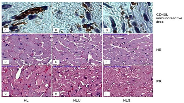
Fig 1. Photomicrograph of transverse histological sections of the left ventricle of mice showing the immunoreactive area for CD40L, cardiomyocyte diameter, and collagen deposition (marked in red).
Group HL: received hyperlipid ration; Group HLU: received hyperlipid feed and grape juice; Group HLS: received hyperlipidic ration and simvastatin.

